-
Reagents
- Flow Cytometry Reagents
-
Western Blotting and Molecular Reagents
-
Flow Cytometry Reagents
- Immunoassay Reagents
- Single Cell Multiomics Reagents
-
Cell Preparation
-
Functional Assays
-
Microscopy and Imaging Reagents
- Western Blotting And Molecular Reagents
- Cell Preparation Separation Reagents
- Functional Cell Based Reagents
- Microscopy Imaging Reagents
- Single Cell Multiomics Reagents
- Single Cell Multinomics Reagents
-
Protocols
- BSB Protocol
-
Setting Compensation Multicolor Flow
-
Tissues Section Stain
-
Immunomicroscopy
-
Immunohistochemical
-
Immunofluorescence
-
Frozen Tissue
-
Parafin Sections
-
Fix Perm Kits
-
Protocol Direct Immunofluorscence Staining
-
Uses of Fc Block
-
Stain Lyse Wash
-
Stain Lyse No Wash
-
Mouse Splenocytes
-
Mouse Rat Leukocytes
-
Isotype Control
-
Indirect Staining Mononuclear Cells
-
Immunopurification
-
Human PBMCs
-
Human Whole Blood Samples
-
Escapee Phenomenon
-
Agarose Conjugates
-
Anti Phosphotyrosine Biotin Conjugates
-
Soluble Antibodies
-
Rabbit Polyclonal Antibodies
-
Monocloncal Antibodies
-
Horseradish Peroxidase
-
Certified Reagents
-
Biotinylated Antibodies
-
Agarose Conjugates X712261
-
Surface Staining
-
Platelet Activation
-
Intracellular Staining
-
Indirect Immunofluorescence
-
Mouse Ige
-
Cytokine Elisa
-
Induction Fas
-
Induction Dx2
-
Apoptosis By Treatment Staurosporine
-
Cell Death
-
Apo Brdu
-
Apo Direct
-
Human Cyclins
-
Detection Ki 67
-
Brdu Detection
-
Targeted mRNA Protocols
-
WTA Protocols
-
360040667732 Protocols
-
360023293831 AbSeq Protocols
-
360039007471 VDJ CDR3 Protocols
-
Annexin V Staining Protocol
-
Western Blotting with Horseradish Peroxidase Conjugates or Alkaline Phosphatase Conjugates
-
Tissue Preparation for Surface Antigen Staining
-
Account Support
-
Account FAQs
- Account FAQ Answer 1
- Account FAQ Answer 2
- Account FAQ Answer 3
- Account FAQ Answer 4
- Account FAQ Answer 5
- Account FAQ Answer 6
- Account FAQ Answer 7
- Account FAQ Answer 8
- Account FAQ Answer 9
- Account FAQ Answer 10
- Account FAQ Answer 11
- Account FAQ Answer 12
- Account FAQ Answer 13
- Account FAQ Answer 14
- Account FAQ Answer 15
- Account FAQ Answer 16
- Account FAQ Answer 21
- Create Account
- Manage Account Settings
-
PrivacyPolicy
-
Terms and Conditions
-
Account FAQs
-
- Account FAQ Answer 1
- Account FAQ Answer 2
- Account FAQ Answer 3
- Account FAQ Answer 4
- Account FAQ Answer 5
- Account FAQ Answer 6
- Account FAQ Answer 7
- Account FAQ Answer 8
- Account FAQ Answer 9
- Account FAQ Answer 10
- Account FAQ Answer 11
- Account FAQ Answer 12
- Account FAQ Answer 13
- Account FAQ Answer 14
- Account FAQ Answer 15
- Account FAQ Answer 16
- Account FAQ Answer 21
- Korea (Korea)
- 국가 / 언어 변경
Old Browser
Overview
BD FACSChorus™ Software simplifies sorting by using a streamlined workflow that eliminates manual setup and monitoring so that you can focus on the science.
The software guides you through the entire cell sorting process by using advanced automation technology that improves throughput and minimizes downtime to promote experimental success.
Features
Features include:
- Automated setup of laser delay, drop delay and side streams
- An interface that leads you through workflows
- On-screen instructions
- Index sorting acquisition and analysis
- Default plots and sort gates
- Prevention of tube overflow
- Automatically turned on deflection plates (only when necessary) to guard operator safety
- FCS 3.1 compatibility
- Proven BD FACS™ Accudrop and BD® Sweet Spot technology
Applications
HEK-293 cells were transfected with 0.25 μg of pAcGFP (Clontech) vector and analyzed for the expression of GFP. Cells expressing variable levels of GFP could be clearly resolved. DAPI was used for exclusion of dead cells. Gates were drawn to identify and sort cells expressing low and high levels of GFP at 1,000 events/second in purity mode. Post-sort analysis revealed purity >99% for both sorted populations.
HEK-293 cells were fixed with 70% ethanol and stained with 0.5 μg/mL of DAPI.
(A) Population hierarchy. Area, height and width parameters for side scatter, forward scatter and DAPI were sequentially used to exclude doublets and cell aggregates. The gating strategy used in this experiment may have underestimated or excluded mitotic figures.
(B) DNA content analysis. Cells within the DAPI singlet gate showed DNA content between 2N and 4N, thus confirming exclusion of >4N doublets or aggregates.
(C) Post-sort validation of single-cell deposition. Cells within the DAPI singlet gate were sorted into three 96-multiwell plates with 99.7% single-cell deposition efficiency. Microscopy was used to confirm the presence of one cell per well. A representative image of a well containing one cell is shown.
Peripheral blood mononuclear cells were isolated from a healthy donor and stained with a cocktail of surface markers (CD3, CD4, CD25, CD127 and CD45RA) for the detection and purification of Treg subsets. Lymphocytes and singlets were first gated based on light scatter properties followed by gating of CD3+CD4+ T cells (not shown). Tregs were then identified as CD127low/neg CD25high. From the Treg gate, CD45RA+ naïve and CD45RA– memory Tregs were identified and sorted at 5,000 events/sec in purity mode. Post-sort analysis revealed homogenous populations of memory and naïve Tregs. Purified cells were then stained for additional surface markers (CD31, CD39 and CD15s) for immunophenotyping. CD31+ recent thymic emigrants (RTEs) were detected within CD45RA+ naïve Tregs, whereas highly activated Tregs were detected within CD45RA– memory Tregs.
Splenocytes from BALB/c mice were isolated following mechanic mincing and enzymatic digestion with collagenase.
(A) The BD IMag™ Mouse Dendritic Cell Enrichment Set was used for bulk selection, resulting in 35- and 29-fold enrichment of CD8α+ and CD8α– subsets of MHC II+CD11chigh conventional dendritic cells, respectively.
(B) Following bulk selection, CD8α+ and CD8α– subsets were sorted at 1,000 events/second in purity mode. Post-sort analysis revealed purity >99% for both sorted subsets.
- Brochure
- Quick Reference Guide
- Training
- BD FACSChorus™ FC Bead Lot Updater
개요
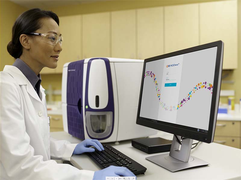
BD FACSChorus ™ Software는 과학적 연구에 집중할 수 있도록 수동 설정 및 모니터링이 필요 없는 간소화된 워크플로우를 사용하여 분류(sorting)를 단순화합니다.
이 소프트웨어는 처리량을 개선하고 가동중지시간을 줄이는 고급 자동화 기술을 이용하여 전체 세포분류 프로세스를 안내하고 실험을 성공적으로 이끕니다.
특징
BD FACSChorus ™ 소프트웨어 설계는 직관적이고 사용하기 쉽기 때문에 초보 사용자도 분류(sorting) 작업에 빠르게 적응할 수 있습니다.
기능:
- Laser delay, drop delay, side stream의 자동설정
- 워크플로우를 안내하는 인터페이스
- 화면에 표시되는 지침
- 인덱스 정렬 획득 및 분석
- 디폴트 plot 및 sort gate
- 튜브overflow방지
- 작업자 안전 보호를 위한 deflection plate(필요한 경우에만) 자동켜짐
- FCS 3.1 호환성
- 입증된 BD FACS™ Accudrop 및 BD® Sweet Spot 기술
응용분야
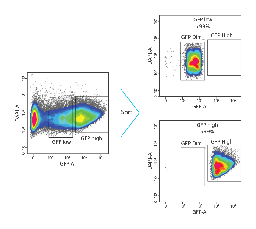
HEK-293 세포를 0.25 μg의 pAcGFP (Clontech) 벡터로 transfection시키고 GFP의 발현에 대해 분석했습니다. 다양한 수준의 GFP를 발현하는 세포는 명확하게 구분 될 수 있습니다. DAPI는 죽은 세포를 배제하기 위해 사용되었습니다. Purity 모드에서 초당 1,000 개 event에서 낮은 수준과 높은 수준의 GFP를 발현하는 세포를 식별하고 분류하기 위해 gate를 그렸습니다. 정렬 후 분석결과 두 집단 모두에서 순도가 99 %를 초과하는 것으로 나타났습니다.
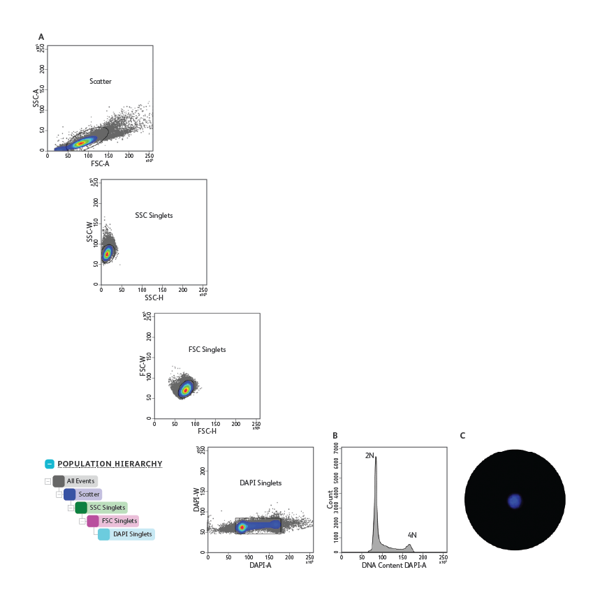
HEK-293 세포를 70 % 에탄올로 고정하고 0.5μg / mL의 DAPI로 염색했습니다.
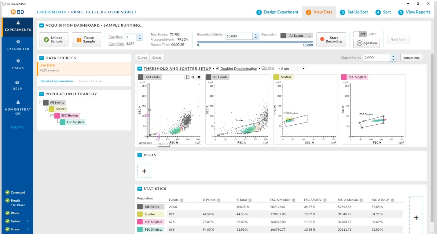
(A) 집단 계층. SSC, FSC 및 DAPI에 대한 A, H 및 W parameter를 순차적으로 사용하여 doublet 및 세포 응집체를 제외했습니다. 이 실험에 사용된 gating 전략은 유사 분열 수치를 과소 평가하거나 배제했을 가능성이 있습니다.
(B) DNA content 분석. DAPI singlet gate 내의 세포는 2N과 4N 사이의 DNA content를 보여주어> 4N doublet 또는 응집체의 배제를 확인했습니다.
(C) Single-cell 증착 정렬 후 검증. DAPI singlet gate의 세포는 99.7 %의 single-cells증착 효율로 3 개의 96 -multiwell plates로 분류되었습니다. 현미경을 사용하여 well당 하나의 세포가 존재함을 확인했습니다. 하나의 세포를 포함하는well의 대표 이미지를 표시했습니다.
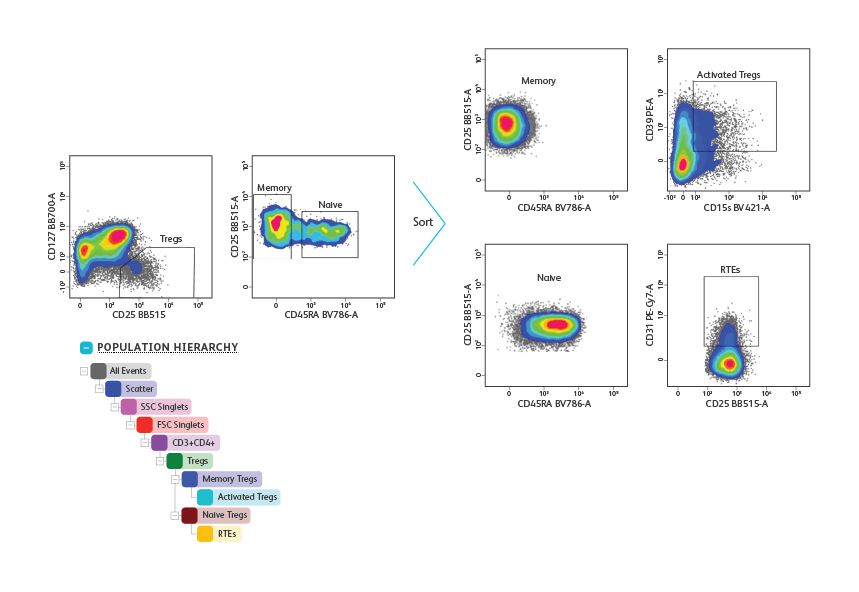
건강한 기증자로부터 얻은 말초 혈액 단핵 세포를 분리하고 Treg subset 의 측정 및 정제를 위해 표면 marker cocktail(CD3, CD4, CD25, CD127 및 CD45RA)로 염색했습니다. 림프구 및 singlet은 우선 FSC,SSC 특성을 기반으로 gating 된 다음 CD3 + CD4 + T cell로 gating(표시되지 않음) 했습니다. Tregs 는CD127low/neg CD25high로 식별되었습니다. Treg gate에서 CD45RA + naïve 및 CD45RA– memory Tregs를 식별하고 purity 모드에서 초당 5,000 개 이벤트로 분류했습니다. 정렬 후 분석 결과 memory와 naïve Treg의 동종 집단이 밝혀졌습니다. 이어서 정제된 세포를 면역 표현형을 위한 추가 표면 marker(CD31, CD39 및 CD15)에 대해 염색했습니다. CD31 + recent thymic emigrants (RTEs)은 CD45RA + naive Tregs 내에서 측정된 반면, 고도로 활성화된 Tregs는 CD45RA- memory Tregs 내에서 측정되었습니다.
BALB/c 마우스 비장 세포는 collagenase를 사용한 기계적 mincing 및 효소적 소화 후에 분리되었습니다.
(A) BD IMag™ Mouse Dendritic Cell Enrichment Set 를 bulk 선택에 사용하여 MHC II + CD11chigh 기존 수지상 세포의 CD8α + 및 CD8α- subset를 각각 35 배 및 29 배 농축했습니다.
(B) Bulk 선택 후, CD8α + 및 CD8α- subset는 purity 모드에서 초당 1,000 개 event로 분류되었습니다. 정렬 후 분석 결과 두 subset에서 모두 순도가 99 %를 초과하는 것으로 나타났습니다.
- BD FACSChorus™ FC Bead Lot Updater
For Research Use Only. Not for use in diagnostic or therapeutic procedures.
23-23320-00
Report a Site Issue
This form is intended to help us improve our website experience. For other support, please visit our Contact Us page.