-
Reagents
- Flow Cytometry Reagents
-
Western Blotting and Molecular Reagents
-
Flow Cytometry Reagents
- Immunoassay Reagents
- Single Cell Multiomics Reagents
-
Cell Preparation
-
Functional Assays
-
Microscopy and Imaging Reagents
- Western Blotting And Molecular Reagents
- Cell Preparation Separation Reagents
- Functional Cell Based Reagents
- Microscopy Imaging Reagents
- Single Cell Multiomics Reagents
- Single Cell Multinomics Reagents
-
Protocols
- BSB Protocol
-
Setting Compensation Multicolor Flow
-
Tissues Section Stain
-
Immunomicroscopy
-
Immunohistochemical
-
Immunofluorescence
-
Frozen Tissue
-
Parafin Sections
-
Fix Perm Kits
-
Protocol Direct Immunofluorscence Staining
-
Uses of Fc Block
-
Stain Lyse Wash
-
Stain Lyse No Wash
-
Mouse Splenocytes
-
Mouse Rat Leukocytes
-
Isotype Control
-
Indirect Staining Mononuclear Cells
-
Immunopurification
-
Human PBMCs
-
Human Whole Blood Samples
-
Escapee Phenomenon
-
Agarose Conjugates
-
Anti Phosphotyrosine Biotin Conjugates
-
Soluble Antibodies
-
Rabbit Polyclonal Antibodies
-
Monocloncal Antibodies
-
Horseradish Peroxidase
-
Certified Reagents
-
Biotinylated Antibodies
-
Agarose Conjugates X712261
-
Surface Staining
-
Platelet Activation
-
Intracellular Staining
-
Indirect Immunofluorescence
-
Mouse Ige
-
Cytokine Elisa
-
Induction Fas
-
Induction Dx2
-
Apoptosis By Treatment Staurosporine
-
Cell Death
-
Apo Brdu
-
Apo Direct
-
Human Cyclins
-
Detection Ki 67
-
Brdu Detection
-
Targeted mRNA Protocols
-
WTA Protocols
-
360040667732 Protocols
-
360023293831 AbSeq Protocols
-
360039007471 VDJ CDR3 Protocols
-
Annexin V Staining Protocol
-
Western Blotting with Horseradish Peroxidase Conjugates or Alkaline Phosphatase Conjugates
-
Tissue Preparation for Surface Antigen Staining
-
Account Support
-
Account FAQs
- Account FAQ Answer 1
- Account FAQ Answer 2
- Account FAQ Answer 3
- Account FAQ Answer 4
- Account FAQ Answer 5
- Account FAQ Answer 6
- Account FAQ Answer 7
- Account FAQ Answer 8
- Account FAQ Answer 9
- Account FAQ Answer 10
- Account FAQ Answer 11
- Account FAQ Answer 12
- Account FAQ Answer 13
- Account FAQ Answer 14
- Account FAQ Answer 15
- Account FAQ Answer 16
- Account FAQ Answer 21
- Create Account
- Manage Account Settings
-
PrivacyPolicy
-
Terms and Conditions
-
Account FAQs
-
- Account FAQ Answer 1
- Account FAQ Answer 2
- Account FAQ Answer 3
- Account FAQ Answer 4
- Account FAQ Answer 5
- Account FAQ Answer 6
- Account FAQ Answer 7
- Account FAQ Answer 8
- Account FAQ Answer 9
- Account FAQ Answer 10
- Account FAQ Answer 11
- Account FAQ Answer 12
- Account FAQ Answer 13
- Account FAQ Answer 14
- Account FAQ Answer 15
- Account FAQ Answer 16
- Account FAQ Answer 21
- Korea (Korea)
- 국가 / 언어 변경
Old Browser
Flow cytometric analysis of CD4 expression on human peripheral blood lymphocytes. Human whole blood was stained with either BD Horizon™ APC-R700 Mouse IgG1, κ Isotype Control (Cat. No. 564974; dashed line histogram) or BD Horizon APC-R700 Mouse Anti-Human CD4 antibody (Cat. No. 564975/564976; solid line histogram). The erythrocytes were lysed with BD FACS™ Lysing Solution (Cat. No. 349202). The fluorescence histogram showing CD4 expression (or Ig Isotype control staining) was derived from gated events with the forward and side light-scatter characteristics of intact lymphocytes. Flow cytometric analysis was performed using a BD™ LSR II Flow Cytometer System.
BD Horizon™ APC-R700 Mouse Anti-Human CD4
Regulatory Status Legend
Any use of products other than the permitted use without the express written authorization of Becton, Dickinson and Company is strictly prohibited.
Preparation And Storage
Product Notices
- This reagent has been pre-diluted for use at the recommended Volume per Test. We typically use 1 × 10^6 cells in a 100-µl experimental sample (a test).
- An isotype control should be used at the same concentration as the antibody of interest.
- Source of all serum proteins is from USDA inspected abattoirs located in the United States.
- Caution: Sodium azide yields highly toxic hydrazoic acid under acidic conditions. Dilute azide compounds in running water before discarding to avoid accumulation of potentially explosive deposits in plumbing.
- Alexa Fluor® is a registered trademark of Molecular Probes, Inc., Eugene, OR.
- For fluorochrome spectra and suitable instrument settings, please refer to our Multicolor Flow Cytometry web page at www.bdbiosciences.com/colors.
- Please refer to www.bdbiosciences.com/us/s/resources for technical protocols.
Companion Products
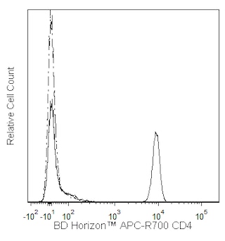
The RPA-T4 monoclonal antibody specifically binds to CD4, a 59 kDa single-chain transmembrane glycoprotein that is expressed on T-helper/inducer cell populations. CD4 is also expressed on thymocyte subsets and at lower levels on monocytes and macrophages. CD4 functions as a co-receptor in MHC class II-restricted antigen-induced T cell activation and as a receptor for human immunodeficiency viruses (HIV). This antibody binds to the D1 domain (CDR1 and CDR3 epitopes) of the CD4 antigen and reacts with approximately 80% of thymocytes and 45% of peripheral blood lymphocytes. RPA-T4 is capable of blocking HIV-1, gp120, and inhibits syncytium formation.
This antibody was conjugated to BD Horizon APC-R700, which has been developed exclusively by BD Biosciences as a better alternative to Alexa Fluor® 700. APC-R700 excites and emits at similar wavelengths to Alexa Fluor® 700 yet exhibits significantly improved brightness. This dye can be excited by the red laser and detected with the same filter set as Alexa Fluor® (eg, 730/45-nm filter).
Development References (3)
-
Knapp W. W. Knapp .. et al., ed. Leucocyte typing IV : white cell differentiation antigens. Oxford New York: Oxford University Press; 1989:1-1182.
-
Piatier-Tonneau D. CD4 workshop panel report. In: Kishimoto T. Tadamitsu Kishimoto .. et al., ed. Leucocyte typing VI : white cell differentiation antigens : proceedings of the sixth international workshop and conference held in Kobe, Japan, 10-14 November 1996. New York: Garland Pub.; 1997:49-54.
-
Schlossman SF. Stuart F. Schlossman .. et al., ed. Leucocyte typing V : white cell differentiation antigens : proceedings of the fifth international workshop and conference held in Boston, USA, 3-7 November, 1993. Oxford: Oxford University Press; 1995.
Please refer to Support Documents for Quality Certificates
Global - Refer to manufacturer's instructions for use and related User Manuals and Technical data sheets before using this products as described
Comparisons, where applicable, are made against older BD Technology, manual methods or are general performance claims. Comparisons are not made against non-BD technologies, unless otherwise noted.
For Research Use Only. Not for use in diagnostic or therapeutic procedures.
23-22944-00
Report a Site Issue
This form is intended to help us improve our website experience. For other support, please visit our Contact Us page.